
Expert insights, surgical technique guides, and medical device updates from industry leaders.
Exploring advancements in joint arthroplasty, trauma management, and spinal systems.
Choosing Cannulated Cancellous Screws in 2026 requires more than comparing diameters, prices, or package labels. The correct choice depends on fracture pattern, bone quality, fragment size, and the planned fixation method. A screw that looks suitable on a catalog page may not fit the patient’s anatomy. Small differences in thread length can affect compression, purchase, and joint safety.
Clinical experience remains important. However, experience should support, not replace, current evidence and careful imaging. Surgeons should review radiographs and, when appropriate, CT scans before selecting screw length and diameter. They should also confirm guidewire compatibility, thread design, head profile, material, and available instrumentation. Manufacturer instructions for use deserve close attention. So do validated mechanical data and peer-reviewed orthopedic research. These details become practical in the operating room, where a smooth guidewire, clear depth measurement, and stable insertion sequence can prevent avoidable problems.
No selection guide is perfect. Patient anatomy varies. Bone quality can surprise the surgical team. Even a well-planned choice may require adjustment during fixation. This article examines how to evaluate Cannulated Cancellous Screws for modern orthopedic procedures, while recognizing the limits of general recommendations. It focuses on informed comparison, patient-specific planning, and responsible clinical judgment. The goal is not to promote one universal screw. It is to help readers ask better questions before choosing a device. That approach is slower, but often safer.
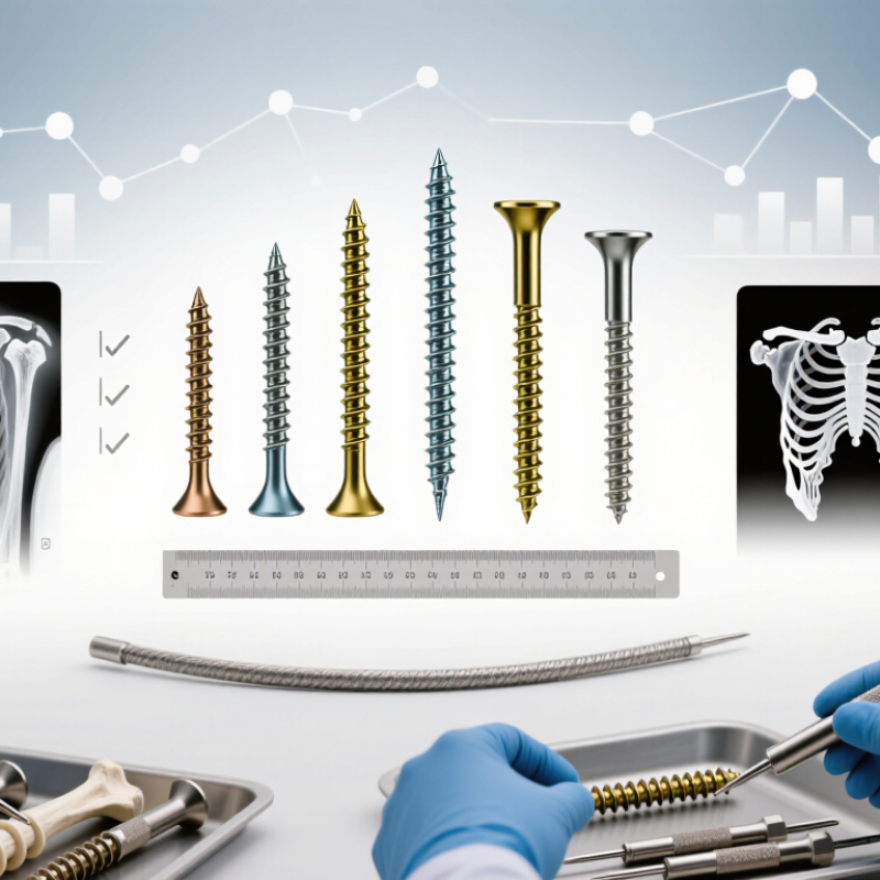
Cannulated cancellous screws combine a hollow shaft with cancellous threads. This design allows guidewire placement before drilling and insertion. The guidewire can improve accuracy near a joint surface. It also helps control screw trajectory through small surgical exposures.
Selection starts with fracture pattern, bone quality, and the required compression. A larger diameter may improve purchase, but it can weaken a narrow fragment. Thread length matters. Threads should cross the fracture sufficiently without entering the joint. Partially threaded screws can create interfragmentary compression. Fully threaded screws may better maintain position when compression is undesirable. A washer can distribute load in softer bone. It may also prevent the screw head from sinking.
The surgeon should confirm screw length with calibrated imaging, not estimation alone. Measure twice. Small errors matter. The cannulation must match the guidewire and compatible instruments. Poor wire control can cause inaccurate drilling or joint penetration. In osteoporotic bone, fixation may need multiple screws, wider spacing, or a different construct. These choices depend on stability goals and the patient’s anatomy.
A common mistake is choosing diameter before understanding the fracture. That sequence feels efficient, but it can produce poor purchase. Another concern is relying on one radiographic view. Multiple views, careful reduction checks, and postoperative assessment provide stronger evidence. Training, validated instruments, and local surgical protocols remain essential, because screw design cannot compensate for an unstable reduction or unsuitable placement.
How to Choose Cannulated Cancellous Screws in 2026?
Choosing a cannulated cancellous screw begins with bone anatomy, not a catalogue chart. Measure the available corridor on thin-slice imaging and confirm it during guidewire placement. Screw diameter should provide reliable purchase without splitting the fragment. In small epiphyseal bone, a slimmer profile may protect cartilage and nearby structures. In stronger metaphyseal bone, a larger diameter can improve thread engagement. The balance is delicate.
Length must support the far fragment while avoiding joint penetration. A screw that is too short may lose fixation. One that is too long can irritate soft tissue or damage the articular surface. Thread length also matters. Partially threaded screws can create interfragmentary compression when the threads cross the fracture line. Fully threaded options may be preferable when controlled positioning matters more than compression. Check the measurement twice.
Fracture pattern should guide the screw arrangement. A simple transverse fracture may accept parallel screws placed perpendicular to the fracture plane. Oblique, vertical, or comminuted patterns often require adjusted trajectories and stronger purchase zones. Poor bone quality may call for washers, multiple screws, or a different fixation strategy. I have seen planning focus too heavily on screw diameter while ignoring entry angle. That mistake is easy to repeat. Surgical judgment, fluoroscopic confirmation, and the patient’s anatomy must remain connected throughout the procedure.
Choosing cannulated cancellous screws in 2026 requires more than matching a catalog size. Thread type should reflect bone quality, fracture pattern, and the desired compression. Partial threads can create interfragmentary compression when the threads remain outside the fracture line. Fully threaded screws may offer better control against shortening. The choice is not automatic. Confirm it under fluoroscopy.
Diameter affects strength, insertion space, and resistance to cutout. A larger screw may improve purchase in suitable bone, but it can weaken a small fragment or crowd nearby implants. Measure the guidewire position carefully. Measure twice. Length should allow adequate thread engagement without penetrating the far cortex or joint surface. A screw that is too short may lose fixation. One that is too long can create a serious mechanical problem.
Material selection also deserves attention. Titanium alloys may reduce imaging artifacts and offer useful biocompatibility. Stainless steel can provide high strength and familiar handling. Consider fatigue resistance, corrosion compatibility, and whether mixed metals are appropriate in the construct. Surgical experience shows that small measurement errors can change the result. Still, anatomy varies, and no sizing rule replaces direct assessment, imaging, and the treating surgeon’s judgment. The selection process should be documented, reviewed, and adjusted when intraoperative findings disagree with the plan.
Selecting thread type, diameter, length, and material for cancellous bone fixation
Partially threaded screws can provide interfragmentary compression when the thread does not engage the near fragment. Fully threaded screws are commonly selected for positional fixation or when compression is not required.
Common cannulated cancellous screw families include approximately 4.0 mm, 6.5 mm, and 7.3 mm diameters. Select the smallest diameter that provides adequate purchase while preserving bone.
Measure the prepared tract and choose a length that achieves the intended thread position without penetrating the far cortex, joint, or nearby soft tissue.
Stainless steel and titanium alloys are commonly used implant materials. Material choice should consider strength, imaging requirements, allergy history, and compatibility with the selected system.
The chart shows representative length bands commonly encountered with major cancellous screw diameter families. Actual availability varies by system and clinical indication; confirm dimensions using surgical planning, imaging, and the applicable device instructions.
Choosing cannulated cancellous screws in 2026 requires more than matching diameter and length. Surgical technique and instrument compatibility should guide the decision. Confirm that the guidewire, drill sleeve, depth gauge, tap, and screwdriver share the same system specifications. A small mismatch can cause poor engagement or inaccurate measurement. Keep the guidewire visible. Protect the far cortex.
Clinical teams should assess bone quality, fracture pattern, thread length, and required compression. A partially threaded screw may provide controlled interfragmentary compression, while a fully threaded option may better preserve position. Fluoroscopy should confirm the entry point, screw trajectory, and final depth in multiple views. Avoid relying on one image. It can mislead.
Safety checks deserve equal attention. The WHO Global Guidelines for the Prevention of Surgical Site Infection report infection rates of about 2–5% in high-income settings and higher rates in lower-resource settings. Sterile handling, validated reprocessing, and minimized operating time remain essential. Inspect cannulated instruments for blocked channels or damaged tips before use. A 2022 systematic review in Injury also emphasized that fixation outcomes depend on fracture stability, reduction quality, and implant positioning, not screw selection alone. That matters.
In practice, a checklist can still miss guidewire migration or hidden instrument wear. Reassess the workflow. Record lot details, imaging findings, and any intraoperative difficulty for future review. Use current surgical instructions and local regulatory requirements.
Choosing cannulated cancellous screws in 2026 requires more than comparing thread length or package price. Clinical evidence should guide the decision. Look for comparative studies reporting union rates, loss of reduction, screw migration, and reoperation. Patient factors matter. Bone quality, fracture pattern, soft-tissue condition, and expected loading can change the risk profile. A screw that performs well in a young cohort may not suit osteoporotic bone. Real-world follow-up is often less tidy than trial data.
Standards offer a practical safety filter. Procurement teams should verify documented mechanical testing, material composition, corrosion resistance, sterility, and manufacturing traceability. ASTM F543 and relevant ISO requirements may support evaluation, but applicable rules differ by market. Ask for current conformity documents, validated cannulation dimensions, and instrument compatibility. Do not treat a certificate as clinical proof. It is only one layer. Surgeon feedback and retrieval findings can expose problems that laboratory testing misses. Small details matter: guidewire fit, fluoroscopic visibility, and consistent head seating.
Cost analysis should include the full episode of care. Add the screw, delivery set, imaging time, operating-room minutes, staff training, inventory loss, and possible revision surgery. A cheaper implant may become expensive when insertion is slow or instruments bend. Yet higher price alone does not prove better outcomes. I would compare cost per successful healing, using local data when possible. That comparison is imperfect. Follow-up may be incomplete, and prices can change. Still, transparent assumptions make the choice more defensible for clinicians, hospitals, and patients.
| Selection Dimension | Common Options | Clinical Relevance | Evidence Interpretation | Standards and Verification Points | Relative Cost Impact | Practical 2026 Decision Rule |
|---|---|---|---|---|---|---|
| Screw diameter | Approximately 4.0 mm, 6.5 mm, and 7.3 mm systems are commonly encountered; available sizes vary by indication and anatomy. | Larger diameters generally provide greater core and pull-out capacity but require adequate bone stock and a safe insertion corridor. | Biomechanical advantages of larger screws do not automatically translate into superior patient outcomes. Clinical choice remains anatomy- and fracture-specific. | Confirm dimensional tolerances, fatigue performance, drive geometry, and compatibility with the system’s instruments. ASTM F543 is commonly used for metallic bone-screw mechanical testing. | Medium | Use the smallest diameter that provides the required fixation strength without compromising reduction or surrounding structures. |
| Thread configuration | Partially threaded or fully threaded; thread lengths selected according to fracture pattern and intended compression. | A partially threaded screw can generate interfragmentary compression when the thread does not engage the near fragment. A fully threaded screw is generally selected when compression is not the primary objective or when controlled fixation is needed. | Compression depends on fracture geometry, screw position, reduction, and technique; thread type alone is not a substitute for accurate placement. | Verify thread profile, pitch, thread-length accuracy, and documented mechanical testing. The implant must be used with the validated drill, guidewire, and countersink sequence. | Low–Medium | Choose partial thread for intentional lag compression when clinically appropriate; choose full thread when maintaining position is more important than compression. |
| Cannulation and guidewire control | Cannulated designs permit guidewire-assisted positioning; guidewire diameter and screw inner diameter must match. | Cannulation can improve trajectory control and facilitate minimally invasive placement, but it introduces risks such as guidewire advancement, loss of purchase, and retained or broken-wire complications. | The main clinical benefit is procedural accuracy and repeatability; evidence does not establish that cannulation alone improves every fracture-healing outcome. | Check guidewire compatibility, cannula patency, radiographic visibility, end-stop design, and instructions for preventing guidewire over-advancement. | Medium | Select a cannulated system when accurate trajectory control or minimally invasive insertion provides a clear procedural advantage. |
| Head and drive design | Countersunk, partially buried, or low-profile heads; commonly paired with a hexagonal or similar internal drive. | Head prominence, soft-tissue irritation, insertion torque, and risk of drive stripping are affected by head and recess geometry. | Low-profile placement may reduce prominence-related symptoms, but the benefit depends on anatomical location and correct countersinking. | ISO 5835 covers dimensions for certain metallic bone screws with a hexagonal drive connection and spherical undersurface. Confirm that the exact implant design falls within the applicable standard. | Low | Prioritize secure torque transfer and appropriate soft-tissue clearance over an unnecessarily small head. |
| Washer availability | Optional washers selected according to screw diameter and local bone quality. | A washer can distribute load over a larger cortical surface and may reduce the risk of screw-head penetration in weak or thin bone. | The value is most situation-dependent; washers may be unnecessary in dense bone and can increase prominence or soft-tissue irritation. | Verify washer–head seating, material compatibility, dimensional fit, and whether the washer is included in the validated construct testing. | Low | Add a washer when load distribution is clinically justified, particularly in thin cortical bone or selected osteoporotic applications. |
| Material and surface | Common implant materials include titanium alloys and implant-grade stainless steels; surface finish may be machined, polished, or otherwise specified by the manufacturer. | Material affects strength, elastic modulus, imaging behavior, corrosion resistance, and compatibility with other implanted metals. | Clinical outcomes are influenced more strongly by indication, reduction, fixation strategy, and patient factors than by material selection alone. | Review material designation, corrosion and galvanic-compatibility data, sterilization status, and biological evaluation under the ISO 10993 framework where applicable. | Medium | Select a documented implant-grade material and avoid unvalidated mixing of dissimilar metals within the construct. |
| Bone quality | Normal, osteopenic, or osteoporotic bone; local cortical thickness may differ substantially from overall bone-density classification. | Poor bone quality increases the risk of pull-out, loss of compression, cut-through, and fixation failure. | A larger screw, washer, altered trajectory, or supplemental fixation may improve construct security, but no single screw feature eliminates biological and mechanical failure risks. | Evaluate implant pull-out and fatigue data in the intended configuration; avoid relying only on nominal diameter or laboratory results from a different construct. | Medium–High | When bone quality is poor, consider load distribution, purchase length, washer use, supplemental fixation, and postoperative protection together. |
| Clinical evidence strength | Evidence commonly consists of biomechanical studies, retrospective cohorts, prospective comparative studies, and systematic reviews. | Outcomes include union, loss of reduction, reoperation, implant irritation, infection, avascular necrosis where relevant, and patient-reported function. | Evidence quality varies by fracture type and anatomical site. Biomechanical superiority should not be presented as proof of superior long-term clinical outcomes. | For procurement, document the indication-specific evidence level, study population, comparator, follow-up duration, and clinically meaningful endpoints. | Indirect cost impact | Give priority to designs supported by evidence relevant to the intended fracture and patient population, not to marketing claims or bench testing alone. |
| Regulatory and quality documentation | Device registration, risk management, manufacturing controls, sterile-barrier validation, and traceability records. | Reliable documentation supports patient safety, recall management, surgical training, and post-market surveillance. | Regulatory clearance or conformity assessment confirms a defined compliance pathway; it does not guarantee superiority over alternative screw designs. | Check applicable jurisdictional requirements, ISO 13485 quality management, risk management under ISO 14971, sterilization validation, and lot traceability. | Medium | Exclude products lacking complete technical files, validated sterile packaging, or clear implant–instrument compatibility information. |
| Total cost of ownership | Implant price, washers, guidewires, trays, sterilization, inventory carrying cost, training, revision risk, and operating-room time. | A lower unit price may be offset by limited size availability, instrument complexity, longer setup time, or higher complication and revision costs. | Economic evidence is highly sensitive to healthcare system, reimbursement rules, case mix, and local operating-room costs. | Use local tender data and a defined costing horizon. Compare equivalent sterile status, accessories, instrumentation, warranty, training, and replacement terms. | High | Compare cost per completed procedure rather than screw price alone; include avoidable revisions, unused inventory, and tray-processing expenses. |
| Recommended minimum specification | A complete range of diameters and lengths, partial and full thread options, compatible guidewires, washers, and validated insertion instruments. | A sufficiently broad range reduces intraoperative substitutions and supports consistent surgical technique. | The most useful evidence is indication-specific and should be linked to the actual implant dimensions and surgical workflow. | Require dimensional drawings, material certificates, mechanical-test reports, biological evaluation, sterilization information, instructions for use, and traceability documentation. | Procurement baseline | Approve the system only when clinical suitability, standards compliance, instrument compatibility, supply continuity, and total procedural cost are evaluated together. |
Start with thin-slice imaging and measure the available bone corridor. Choose enough purchase without splitting the fragment. Small epiphyseal bone may need a slimmer profile. Stronger metaphyseal bone may accept a larger diameter. The balance is delicate.
The screw should support the far fragment without entering the joint. A short screw may lose fixation. A long screw can irritate soft tissue or damage cartilage. Check the measurement twice.
Partially threaded screws can create interfragmentary compression. Their threads should cross the fracture line correctly. Fully threaded screws may suit controlled positioning better. The fracture goal matters more than a simple preference.
A transverse fracture may accept parallel screws perpendicular to the fracture plane. Oblique, vertical, or comminuted fractures need adjusted trajectories. Poor bone quality may require washers or multiple screws. Diameter alone does not solve every problem.
The guidewire, drill sleeve, depth gauge, tap, and screwdriver should share matching specifications. Small mismatches can reduce engagement or distort measurement. Keep the guidewire visible. Protect the far cortex.
Fluoroscopy should confirm the entry point, trajectory, and final depth. Use multiple views. One image can mislead. Recheck guidewire position before advancing the screw. A checklist may still miss migration.
Inspect cannulated instruments for blocked channels or damaged tips. Use sterile handling and validated reprocessing. Minimize unnecessary operating time. Record imaging findings and difficult steps for later review. Perfect workflows are uncommon.
Review studies reporting healing, loss of reduction, migration, and reoperation. Consider bone quality, soft tissue, fracture pattern, and expected loading. Cost should include imaging time, operating-room minutes, training, inventory loss, and possible revision. A lower price does not prove better value.
Request mechanical testing, material information, corrosion data, sterility records, and manufacturing traceability. Confirm cannulation dimensions and instrument compatibility. Standards can support evaluation, but they do not prove clinical success. Surgeon feedback still matters.
Choosing Cannulated Cancellous Screws in 2026 requires a balanced evaluation of design, anatomy, fracture characteristics, surgical workflow, and long-term value. Their hollow structure can support guidewire-assisted placement, while thread configuration, core strength, diameter, and length should be matched to bone quality, fracture location, fragment size, and the required compression. Material selection should also consider biocompatibility, mechanical performance, imaging needs, and the possibility of future removal.
Surgeons should verify that the screws, guidewires, drills, drivers, and measurement tools are fully compatible and suitable for the planned technique. Safe placement depends on accurate reduction, controlled drilling, protection of surrounding structures, and confirmation of screw position. Clinical evidence, applicable quality standards, sterilization requirements, and documented performance can help compare available options. Finally, cost should be assessed alongside reliability, instrument needs, training, inventory management, and overall patient-care value rather than considered as the only deciding factor.